Clinical tip: Metacarpal and metatarsal fractures “spider frame”
This is the first of what I hope will become a collection of occasional posts with clinical tips/techniques based on cases we have seen.
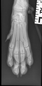
This week we were asked to see a young Springer Spaniel puppy that had injured its foot.
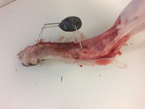
There are a number of ways to treat metacarpal/metatarsal fractures, including conservative management with a bandage/splint or plating. My preference in cats and smaller dogs is to use a “spider frame” – which consists of K-wires placed in an intra-medullary fashion exiting through the skin distally, bent around and incorporated into a lump of “epoxy putty“. This technique was described by Noel Fitzpatrick and I have had good success with it in a number of cases. The procedure is quick and fairly straightforward, and does not require expensive implants compared to the cost of very small plates and screws.
The procedure is performed as follows:
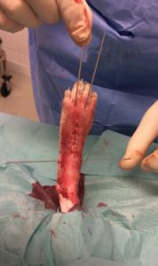
- A dorsal midline incision is made over the third and fourth metacarpal/tarsal bones (unless the injury is asymmetric)
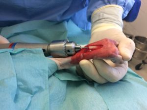
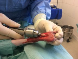
- The fracture site(s) is identified and a suitable sized K-wire is retrograded from the fracture site distally (this can be done with a hand chuck or with a power drill)
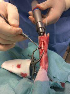
- The pin is exited through the skin at the MCP/MTP joint and withdrawn until the tip of the pin is flush with the fracture end
- The fracture is reduced and the pin is advanced into the proximal part of the metacarpal/tarsal bone
- This is repeated for as many bones as are fractured. It may be sufficient to only treat the two central bones (3 & 4) but I will generally pin all of the fractured bones.
- A pin is placed transversely across the heads of the metacarpals/metatarsals
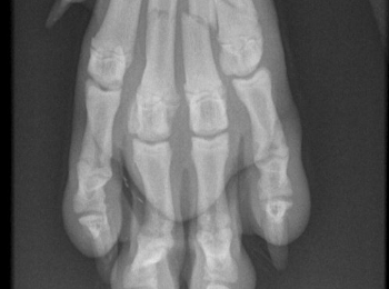
- The skin is closed and radiographs are obtained.
- If the pins are correctly placed then they are bent around and incorporated into epoxy putty
- Radiographs are obtained at approximately 6 weeks to assess healing. The frame is generally removed at some point between 6 & 10 weeks

